Several weeks ago I was perched alongside the Canadian River in Norman, Oklahoma. It was not long after sunset. Surprisingly, I could hear people talking out on the river, though they seemed a ways away. Subsequently, I heard extended rustling in the leaves along the riverbank, which could only be the sounds of a human. Then suddenly there he was, a very white man in the eerie gloaming, some 5′ 7” maybe, standing among the bushes in a manner altogether agnostic as to the presence of any clothing.
“Looks like an insect light,” he cheerfully exclaimed. And it was.
“Ah, yes! A blacklight blue.”
“What are you looking for?”
“Are you an entomologist? I’m trying to catch a nocturnal species of Strepsiptera.”
“Ah, Strepsiptera…” And then he emerged fully from the brush – wearing a bathing suit, by the way – and sat down on a log by the light. “What makes you think there are Strepsiptera here?” And so began a conversation with Ken Hobson, former roommate of a professor of mine at the University of California, Davis, half a continent away.
“I read an article. By Bill Shepard. He caught a bunch of them in mosquito light traps.” 35 years ago, but you go with what you can get. “I’ve got the mosquito board’s by-catch. They’re here, alright. I don’t know about here. But their host is meant to like loamy damp soil. I’m trying to figure out when they fly. I need them live.”
You can ‘fix’ the eyes of an insect caught live. That is, stabilize their form for more detailed analysis. Dead animals have the tendency to dehydrate or to be partially consumed by microorganisms – and quickly. Such occurrences destroy internal structures, and those structures are likely to be very different in the eyes of Strepsiptera.
A brief glimpse into a compound eye
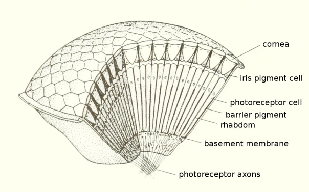
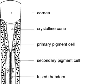
Compound eyes are all about modularity – modularity and repetition with variation. As such, they all contain a number of remarkably similar visual elements called rhabdoms. Each rhabdom sits under its own lens, the outer surfaces of which make up the facets of the compound eye – like the the faces of a minuscule jewel. The lens, its rhabdom, and other support cells are together called an ommatidium [New Latin, from Greek ommat-, omma: eye]. Each ommatidium is a tiny light sensitive organ – a minute eye. But an eye only capable of resolving a single point.1 An individual ommatidium can potentially perceive information about the brightness (intensity), color, and/or polarization of that point. But to get a decent image, you’ve got to pool the output of several of them. The various ways in which ommatidia are amassed to create an organ capable of perceiving form is the art and wonder of vision via compound eyes.


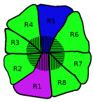
A rhabdom is itself composed of even smaller light absorbing tissues called rhabdomeres. Insect compound eyes typically have six rhabdomeres that are all most responsive to green light and apparently support their exquisite sensitivity to motion. These identical rhabdomeres are generally grouped with two other rhabdomeres that respond to different wavelengths of light – different from green, and usually different from one another too. Those two, along with one or more of the green-absorbing rhabdomeres, have been found to support color vision. Like humans, most insects have trichromatic color vision – color vision that is mediated by three classes of photoreceptors, each responding maximally to a different wavelength of light. For normally sighted humans, those separate peaks are in the blue, green, and red2 ranges of the electromagnetic spectrum3 (EM). These primary colors are combined additively to represent all colors available to the visual system. For trichromatic insects, however, the three peaks are in the UV, blue, and green ranges – they’re shifted to EM radiation having shorter wavelengths. But some insects are tetrachromats, having four primary colors, and thus the prerequisite machinery to see many more colors than humans (many butterflies, for example, and fittingly, a lot of butterflies can see red), and others are dichromats (ants).

Polarization Primer
Polarization is a property of light our eyes are blind to, by and large. (But see Haidinger’s Brush.) The nexus of that is the organization of our photoreceptors. You can think of polarization as an angular vibration every photon has that can give information about what it most recently interacted with. In order to absorb a photon’s energy and begin the process of seeing, a photopigment – the molecules of a photoreceptor that absorb light energy – has to be sufficiently attuned to that photon’s wavelength (energy level) and its polarization state. When photopigments are ordered haphazardly, then light of all different polarization angles can be absorbed with equal likelihood. Since sunlight is “unpolarized”, that is, it contains photons with random polarization angles, and the polarization angle of reflected or refracted light cannot be ascertained for all environments a priori, it generally takes less light to see using photoreceptors that can absorb photons of any polarization. This condition also removes the possibility of perceiving false colors, in case some wavelengths of light are more likely to have some polarization angles than others (depending on what’s been interacted with). That would mean that some wavelengths of light would be more easily absorbed than others – not because there is more light of that wavelength, but instead because of the light’s polarization. That, in turn, could be interpreted as a change in the color of the light, when, in fact, no such change has occurred. On the other hand, polarization sensitivity can provide navigational queues from scattered skylight, and can be the basis for glare reduction, and it may allow one to detect camouflaged objects by means of their polarization properties rather than their cryptic colors. For such reasons, many organisms are polarization sensitive in a part of their eyes, or in specialized secondary eyes.
